Research
Our research links neuroscience to applied fields of sport, physical activity and training.
The goal is to develop specific and personalized training strategies for healthy young and elderly people, patients and athletes on the basis of a thorough understanding of the mechanisms of brain plasticity.
Projects in Research Focus 1 investigate the role of training for brain health and performance from a basic science perspective.
Projects in Research Focus 2 examine how individual cognitive abilities unfold in complex real-world situations.
Our basic and applied science projects are supported by the Deutsche Forschungsgemeinschaft (DFG), Bundesinstitut für Sportwissenschaft (BISp) and the Otto-von-Guericke University (OvGU).
Training-induced Brain Plasticity

Longitudinal neuroimaging research in the past 20 years provided compelling evidence for training-induced changes in brain structure and function. Initial findings of localized grey and white matter changes (Draganski et al., 2004 Nature; Scholz et al., 2009 Nature Neuroscience) set the stage for a wave of human plasticity research that demonstrated brain changes within different settings and through diverse training stimuli. Such findings have great promise for healthy cognitive aging, diseases prevention, rehabilitation and human performance enhancement. In the past years, however, progress in the field has been dampened for both methodological and conceptual reasons.
We believe that further progress will emerge from a better understanding of the functional role and the neurobiological underpinnings of training-induced brain plasticity.
Along these lines, our projects seek to better understand the following main features:
Role of brain structural and functional plasticity in cognitive and motor transfer
Description:
The project aims at establishing a link between behavioral training, brain plasticity and behavioral performance improvements. Using training interventions such as physical exercise (i.e. high-intensity or moderate-intensity exercise) and motor balance learning (i.e. stabilometer task), we identify neural mechanisms of brain function and structure that mediate between training and improved task/transfer performance. The research approach employs longitudinal brain imaging techniques with repeated measurements across training. Key magnetic resonance imaging (MRI) modalities include diffusion-weighted MRI and functional resting-state MRI. In this project, we collaborate with colleagues from the Max-Planck-Institute for Human Cognitive and Brain Sciences in Leipzig (Prof. Arno Villringer). We have recently shown that exercise-induced structural changes in white matter tracts underlying the prefrontal cortex as well as exercise-induced increases in prefrontal cerebral blood flow mediate the beneficial effect of exercise on motor balance learning (Lehmann et al., 2020a).
In another recent study, we showed that exercise-induced increases in functional network connectivity of the dorsal prefrontal cortex mediate the benefit of exercise on sustained visual attention (Lehmann et al., 2020b). These studies demonstrate the transfer potential of training-induced brain plasticity and motivate a closer inspection of the behavioral triggers of brain plasticity and its modulation by age and pathology. We address this new perspective in close collaboration with colleagues from Universities of Lausanne (Prof. Bogdan Draganski) and Fribourg (Prof. Wolfgang Taube) as well as the Medical Faculty at OvGU Magdeburg (C01 of SFB 1436 Neural Resources of Cognition together with Dr. Gabriel Ziegler from the Institute for Cognitive Neurology and Dementia Research).
Our results support current efforts to develop more efficient physical exercise and motor learning paradigms in the context of mental health and cognitive ageing.
Selected publications:
Lehmann, N., Villringer, A. & Taubert, M. (2022).
Priming cardiovascular exercise improves complex motor skill learning by affecting the trajectory of learning-related brain plasticity.
Scientific Reports, doi: 10.1038/s41598-022-05145-7
Lehmann, N., Villringer, A. & Taubert, M. (2020).
Intrinsic Connectivity Changes Mediate the Beneficial Effect of Cardiovascular Exercise on Sustained Visual Attention.
Cerebral Cortex Communications. doi: 10.1093/texcom/tgaa075
Lehmann, N., Villringer, A. & Taubert, M. (2020).
Co-Localized White Matter Plasticity And Increased Cerebral Blood Flow Mediate The Beneficial Effect Of Cardiovascular Exercise On Long-Term Motor Learning.
The Journal of Neuroscience, 12, 2416-29. doi: 10.1523/JNEUROSCI.2310-19.2020.
Woost, L., Bazin, P.L., Taubert, M., Trampel, R., Tardif, C.L., Garthe, A., Kempermann, G., Renner, U., Stalla, G., Ott, D.V.M., Rjosk, V., Obrig, H., Villringer, A., Roggenhofer, E. & Klein, T. (2018).
Physical exercise and spatial training – a longitudinal study of effects on cognition, growth factors, and hippocampal plasticity.
Scientific Reports, 1, 4239. doi: 10.1038/s41598-018-19993-9.
Temporal and spatial dynamics of training-induced brain plasticity
Description:
Improvements in performance result from physiological adaptations of the body. While peripheral adaptations (such as cardiovascular and musculoskeletal adaptations) meet the energetic demands of improved (or sustained) aerobic performance, central adaptations in the brain form the physical basis for an improved information processing and storage during various types of learning. Advancements in neuroimaging technology over the past 30 years have enabled researchers to track functional and structural brain adaptations in the living human brain in response to acute or prolonged periods of motor learning. A better understanding of these central brain adaptations, i.e. its temporal and spatial dynamics during learning, will lead to new intervention strategies in the context of healthy ageing or neurological rehabilitation – much like the understanding of peripheral adaptations has contributed to practical implementations of endurance and strength training methods.
Studies from our lab provided novel evidence for rapid, transient as well as performance-related structural brain changes over the course of motor balance learning (Taubert et al., 2010; Taubert et al., 2016). Learning a new balance task leads to rapid grey matter changes in motor representations of the primary motor cortex (Taubert et al., 2016). After two weeks of practice, grey matter expansions were found in supplementary motor, inferior parietal and dorsolateral prefrontal brain regions along with enhanced functional resting-state connectivity (Taubert et al., 2010, 2011). With further practice, however, these initial structural and functional changes renormalized and returned to baseline levels. This pattern was found although our volunteers further improved their balance performance during practice. We additionally found a steadily evolving and performance-related grey matter expansion across the whole learning period in parts of the frontopolar cortex which was paralleled by steadily-evolving microstructural changes in white matter underlying the frontopolar cortex (Taubert et al., 2010).
This non-linear time course of plasticity with rapid but transient structural brain changes was also found by other groups using other motor learning tasks (e.g. Wenger et al., 2016). The findings can be conceptualized in an expansion-renormalization model of brain plasticity (Wenger et al., 2017). This model offers a new perspective that views brain plasticity as a valuable but temporally limited neural resource for improved (transfer) performance.
In a new project in collaboration with Dr. Gabriel Ziegler from the Institute for Cognitive Neurology and Dementia Research in Magdeburg, we will investigate the temporal and spatial dynamics of training-induced plasticity in the elderly brain. Novel imaging techniques will allow us to quantify trajectories of plasticity in grey and white matter microstructure.
Selected publications:
Taubert, M., Mehnert, J., Pleger, B. & Villringer (2016).
Rapid and Specific Grey Matter Changes Induced by Balance Training.
NeuroImage, 133, 399-407. doi: 10.1016/j.neuroimage.2016.03.017.
Sehm, B., Taubert, M., Conde, V., Weise, D., Classen, J., Dukart, J., Draganski, B., Villringer, A. & Ragert, P. (2014).
Structural brain plasticity in Parkinson’s disease induced by balance training.
Neurobiology of Aging, 1, 232-239. doi: 10.1016/j.neurobiolaging.2013.06.021.
Taubert, M., Lohmann, G., Margulies, D.S., Villringer, A. & Ragert, P. (2011).
Long-term effects of motor training on resting-state networks and its underlying brain structure.
NeuroImage, 4, 1492-1498. doi: 10.1016/j.neuroimage.2011.05.078.
Taubert, M., Draganski, B., Anwander, A., Müller, K., Horstmann, A., Villringer, A. & Ragert, P. (2010).
Dynamic properties of human brain structure: learning-related changes in cortical areas and associated fiber connections.
The Journal of Neuroscience, 30, 11670-11677. doi: 10.1523/JNEUROSCI.2567-10.2010.
Brain tissue properties in health and disease from macro- to microstructural levels
Description:
Magnetic resonance imaging (MRI) is a widely used tool to measure brain function and structure in living human subjects. Advantages of this technique include its non-invasive character and the possibility to measure volunteers repeatedly (i.e. across training). This makes MRI a valuable tool to study the brain and its changes over time at very high spatial resolution. However, most MRI studies in the field of training-induced brain plasticity have used MR sequences and analysis techniques that capture morphometric (shape and size) aspects of grey and white matter.
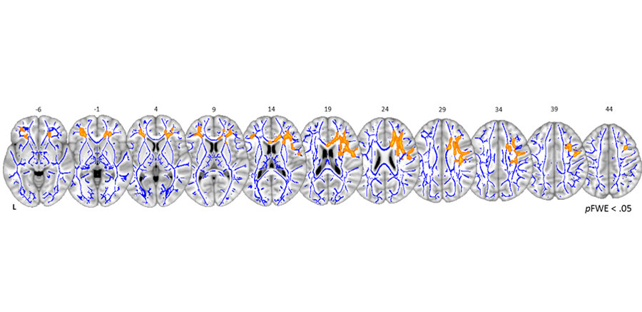
While these studies provided important insights into plastic changes of brain macrostructure, these approaches are of limited value for investigations of biologically-plausible plasticity mechanisms at the microstructural level. To overcome this issue, promising new quantitative MRI protocols have been developed over the past years. These protocols seem to have a higher biological specificity and quantify brain tissue properties that are linked to important histological markers such as myelin or iron. Together with colleagues from the University of Lausanne (Prof. Bogdan Draganski) we were able to show associations between theses brain tissue properties and participants’ age in a sample of approx. 1000 healthy elderly volunteers (Taubert et al., 2020). The results point to a pronounced age-related demyelination in white matter tracts of the prefrontal cortex as well as in grey matter regions of the primary motor cortex.
These patterns were accompanied by an enhanced iron deposition in subcortical regions with advanced years. Whether those age-related signatures contain behaviorally-relevant information or whether and how these maladaptive tissue properties are modifiable through training is presently unclear.
In order to prepare for future longitudinal training studies with this methodology, our lab currently evaluates the reproducibility of those new MRI sequences in collaboration with colleagues from the Medical Faculty in Magdeburg. We particularly focus on the reproducibility of multi-parametric maps and bio-physical modelling of multi-shell diffusion-weighted MRI.
Selected publications:
Aye, N., Lehmann, N., Kaufmann, J., Heinze, H.-J., Düzel, E., Taubert, M. & Ziegler, G. (2022). Test-Retest Reliability of Multi-Parametric Maps (MPM) of Brain Microstructure. NeuroImage, https://doi.org/10.1016/j.neuroimage.2022.119249
Taubert, M., Roggenhofer, E., Melie-Garcia, L., Muller, S., Lehmann, N., Preisig, M., Vollenweider, P., Marques-Vidal, P., Lutti, A., Kherif, F. & Draganski, B. (2020). Converging patterns of aging-associated brain volume loss and tissue microstructure differences. Neurobiology of Aging, 88, 108-18. doi: 10.1016/j.neurobiolaging.2020.01.006.
Lehmann, N., Aye, N., Kaufmann, J., Heinze, H.J., Düzel, E., Ziegler, G. & Taubert, M. (2021).
Longitudinal reproducibility of neurite orientation dispersion and density imaging (NODDI) derived metrics in the white matter. Neuroscience, https://doi.org/10.1016/j.neuroscience.2021.01.005
Cognitive Abilities and Behavior

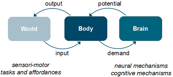
In real-world situations, human behavior is shaped by environmental and situational characteristics as well as individual dispositions of the performer.
An implicit but largely unproven assumption is that training of particular cognitive or motor functions under standardized conditions will benefit behavior under real-world conditions. Previous efforts from experimental scientists have revealed prominent improvements in trained tasks but a limited transfer to other tasks in response to lab-based training interventions. In contrast to this, training methods from sports scientists and/or practitioners from various athletic disciplines are explicitly developed to improve competitive performance under highly challenging conditions (competitive stress, decision-making in limited amount of time, coordinated collective performance, presence of opponents etc.). Thus, individual improvements in training may or may not unfold in real-world situations – be it in sports domains or in other aspects of everyday life.
Our research aims to better characterize individual behavior and individual performance improvements under more complex and challenging conditions than in a usual lab environment.
Much like our projects in Research Focus 1, we will investigate the transfer of training-induced performance improvements to other related tasks as well as performance under different environmental and situational conditions. In our research projects, we consider transfer along a continuum from proximal (conditions more similar to the trained scenario) to more distal (conditions with additional influencing factors) conditions.
Diagnostics and training of individual cognitive abilities in team sports
Description:
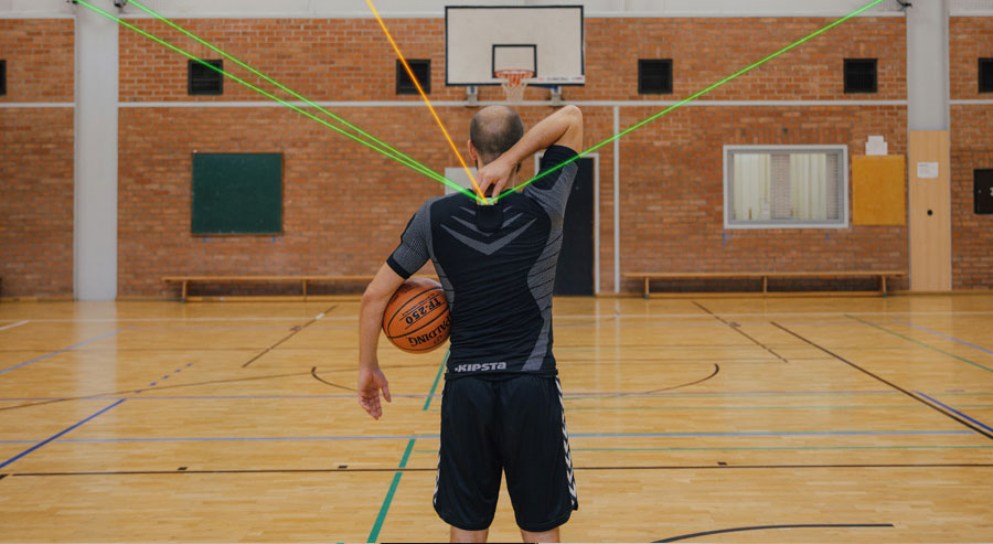
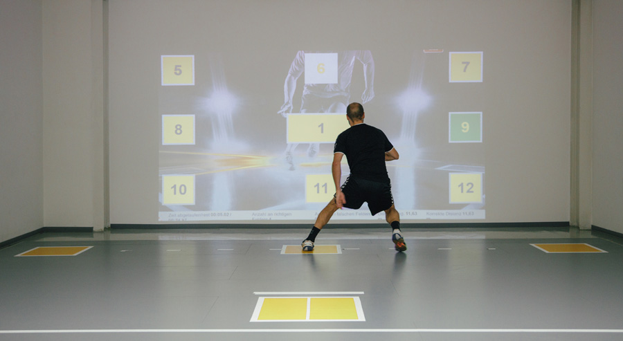
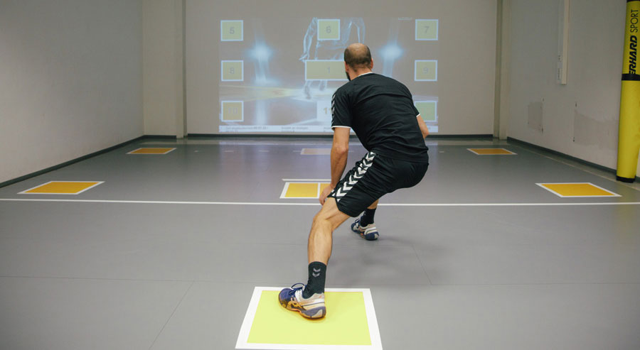
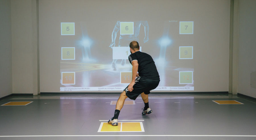
This project develops a test battery to assess an individuals’ ability to predict forthcoming actions of an opponent in team handball. Performance in team sports depends on a variety of individual and collective attributes.
The ability of players to predict the actions of their opponents is of critical relevance for success as it helps f. ex. to (re)act quicker and to appropriately defend opponents attack situations. Previous tests of predictive performance in team sports were largely based on visual stimulation coupled with simple response characteristics (button press in seating or standing position). In our project we evaluate (test reliability and validity) a battery of tests to assess predictive performance under more challenging response characteristics (e.g. specific defensive actions such as short sprints).
Test validity will be evaluated with an expert-novice paradigm in close collaboration with the local handball clubs (e.g. SC Magdeburg). The project is funded by the Bundesinstitut für Sportwissenschaft (BISp). Please see this video for more information.
Publications:
Hinz, M., Lehmann, N., Melcher, K., Aye, N., Radic, V., Wagner, H. & Taubert, M.(2021). Reliability of Perceptual-Cognitive Skills in a Complex, Laboratory-Based Team-Sport Setting. Applied Sciences, https://doi.org/10.3390/app11115203
Hinz, M., Lehmann, N., Aye, N., Melcher, K., Tolentino-Castro, W., Wagner, H. & Taubert, M. (2022). Differences in Decision-Making Behavior between Elite and Amateur Team-Handball Players in a Near-Game Test Situation. Frontiers in Psychology-Movement Science and Sport Psychology, https://doi.org/10.3389/fpsyg.2022.854208
Individual and collective behavior
Description:
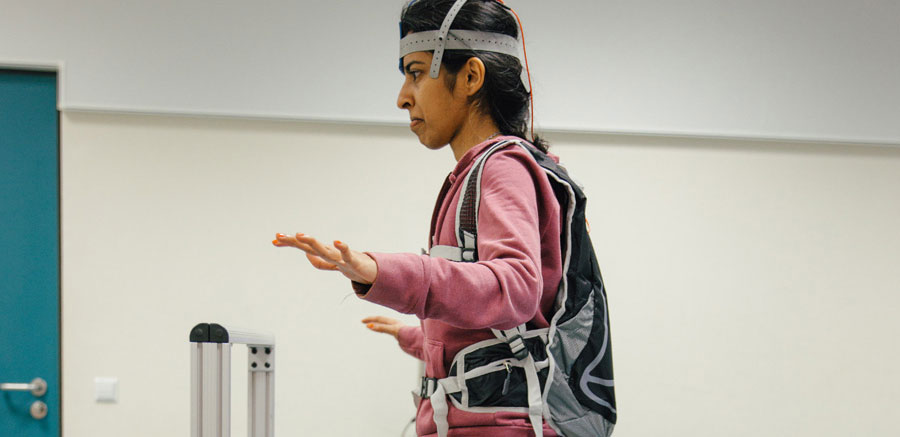
This project aims at investigating the movement of multiple participants within groups using real-time positional tracking methodology. Coordinated group performance is an important component of successful teams.
Assessment of group performance for scientific purposes is so far largely based on subjective ratings or laborious and time-consuming video analyses. Real-time positional tracking systems offer advantages as they record each player’s field position with high temporal and spatial accuracy. Our goal is to infer the cognitive and motor capabilities of individuals and/or groups from positional tracking data under experimental conditions.
We currently validate our tracking system and test new analysis algorithms to quantify group tactical behavior (in close collaboration with Prof. Matthias Kempe from University of Groningen).
Research Facilities
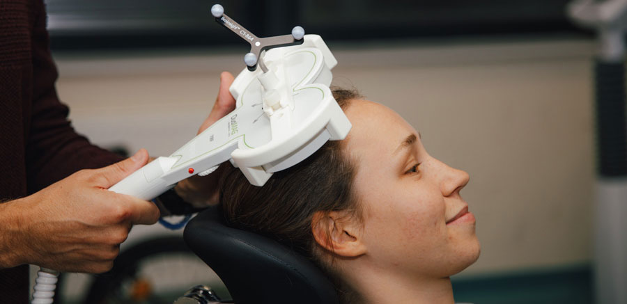
- Transcranial magnetic stimulation (single- and paired pulse; intra- and interhemispheric interactions)
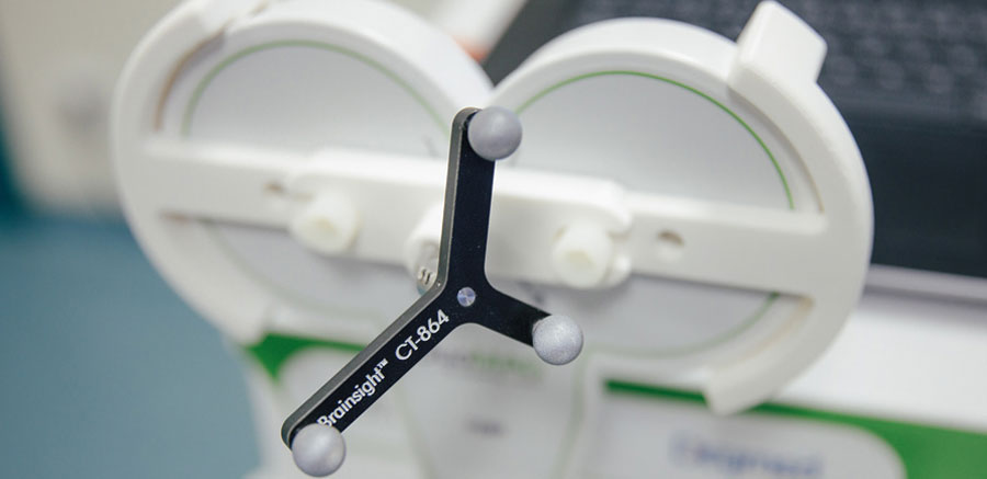
- Brainsight Neuronavigation
- Transcranial electrical stimulation (tDCS, tACS)
- Magnetic Resonance Imaging (MRI) at 3T (at the Dept. Neurology at OvGU)
- Stabilometer (Model 16030, Lafayette Instruments) to study balance learning
- Inverse steering bike to study motor interference and consolidation
- Light beams for locomotor speed assessment
- Bicycle ergometer for performance diagnostics and controlled exercise interventions
- Stationary PC and several laptops for cognitive testing in lab and field environments
- Real-time tracking system for indoor positional data analysis (in cooperation with ST Sportservice)
- Polar Team Pro system for outdoor positional and physiological data analysis
- SpeedCourt system (GolbalSpeed GmbH) for performance diagnostics and training